

NEW PUBLICATIONS UPTO NOV. 21ST 2015
NEW PUBLICATIONS UPTO NOV. 21ST 2015


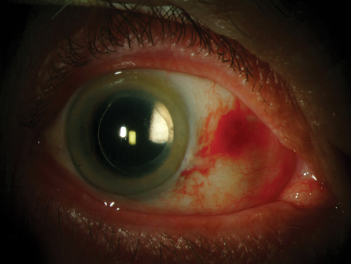
By Josh Johnston, OD, FAAO 11/15/2015 A 63-year-old female presented in the clinic for an eye exam for the first time in five years with a best-corrected visual acuity of 20/40 OD and 20/30 OS, with glare disability of 20/80 OU.
http://www.reviewofcontactlenses.com
|
1.
|
Clin Ophthalmol. 2015 Nov 2;9:2039-2047.
Potvin R1, Makari S1, Rapuano CJ2.
Author information:
Abstract
OBJECTIVE:
To evaluate the evidence in the peer-reviewed literature regarding the use of tear osmolarity as a physiological marker to diagnose, grade severity, and track therapeutic response in dry eye disease (DED). In addition, to review the evidence for the role of tear osmolarity in the pathophysiology of DED and ocular surface disease. METHODS:
A literature review of all publications after the year 2000, which included the keywords "tear osmolarity", was conducted. Relevant articles were graded according to quality of evidence and research, using the University of Michigan Practice Guideline and the Grading of Recommendations Assessment, Development, and Evaluation (GRADE) rating systems. Articles were further categorized by the nature of any reported financial support and by the overall impression they provided related to tear osmolarity. RESULTS:
A total of 164 articles were identified as relevant to the search criteria, although some were editorials, and some were written in a foreign language. Of the total, it was possible to grade 159, and an overall impression was generated for 163. A positive impression of tear osmolarity in DED diagnosis was evident in 72% (117/163) of all articles, with a neutral impression in a further 21% (35/163); 7% had a negative impression. The percentage of positive impressions appeared independent of the quality of research; 73% (38/52) of articles graded high/moderate quality supported the use of tear film osmolarity measurement in DED diagnosis. Impressions were also independent of the source of financial support, with 72% (75/104) of independent studies positive. CONCLUSION:
The literature broadly supports the use of tear film osmolarity as an objective numerical measure for diagnosing, grading severity, and managing treatment of DED. |
| PMID: 26586933 [PubMed - as supplied by publisher] | |
| Similar articles | |
  |
| 2. | Mini Rev Med Chem. 2015 Nov 19. [Epub ahead of print]
Author information:
Abstract
Extensive research during the past three decades has demonstrated the mechanisms by which an imbalance in the redox status of prooxidant/antioxidant reactions in cells with advantage of prooxidant reactions (oxidative stress, OS) can cause peroxidation of nucleic acids, bases, lipids, proteins and carbohydrates, thus resulting in their damage. These actions result in stimulation of signal transduction pathways and activation of transcription factors that can lead to chronic inflammation and cause tissue dysfunction. The most important oxidants are reactive oxygen species (ROS) and reactive nitrogen species (RNS) generated by various metabolic pathways, physical, chemical and biological factors, and pathological conditions. The eye is one of the major target of the ROS/RNS attack due to exposition on several environmental factors like high pressure of oxygen, light exposure, ultraviolet rays, ionizing radiation, chemical pollutants, irritant, and pathogenic microbes, which are able to shift the redox status of a cell towards oxidizing conditions. There is increasing evidence indicating that persistent OS contributes to the development of many ocular diseases. Increases in the accumulation of hydrogen peroxide and markers of the oxidative damage to DNA, lipids, proteins observed in several eye diseases and usage of antioxidants in their treatment and prevention emphasize the involvement of OS pathways. This paper summarizes the present state of knowledge in the involvement of OS in the etiology of non-cancer ocular diseases (dry eye syndrome; corneal and conjunctive diseases; cataract; glaucoma; age-related macular degeneration; retinitis pigmentosa; diabetic retinopathy, autoimmune and inflammatory uveitis) and cancer ocular diseases (melanoma; retinoblastoma; lymphoma). The paper also discusses the potential applications of antioxidants in the prevention of eye diseases and shows a duality of physical exercise actions: protection against the ROS/RNS damage by regular-moderate physical activity and damaging effect through mediation of OS by endurance exercise without adaptable physical training. |
| PMID: 26586128 [PubMed - as supplied by publisher] | |
| Similar articles | |
| 3. | Cornea. 2015 Nov 17. [Epub ahead of print]
Author information:
Abstract
PURPOSE:
Inflammation and squamous metaplasia is a common pathological process that occurs in many ocular surface diseases such as dry eye, Stevens-Johnson syndrome, mucous membrane pemphigoid, and chemical/thermal burns. At present, there is no ideal medicinal treatment for this abnormality. We report herein on an ex vivo conjunctival inflammation and squamous metaplasia model by culturing human conjunctival tissues at an air-liquid interface for up to 8 days to study the effects of minocycline on inflammation and squamous metaplasia. METHODS:
The levels of inflammatory mediators including interleukin-1β, tumor necrosis factor-α, and metalloproteinase-9 in the cultured human conjunctival tissues were determined by enzyme-linked immuno-sorbent assay and real-time polymerase chain reaction. The total and phosphorylated nuclear factor-κB were detected by western blot. Differentiation markers K10, MUC5AC, and Pax6 and proliferation markers Ki67, p63, and K14 were determined by immunofluorescence or immunohistochemical staining. RESULTS:
The results indicated that minocycline inhibited inflammation, decreased the expression of interleukin-1β, tumor necrosis factor-α, and metalloproteinase -9, and squamous metaplasia features such as hyperproliferation and abnormal epidermal differentiation of conjunctival epithelium. CONCLUSIONS:
These findings highlight the possibility that minocycline could be used to treat dry eye and other ocular surface diseases exhibiting epithelial cell inflammation and squamous metaplasia. |
| PMID: 26583279 [PubMed - as supplied by publisher] | |
| Similar articles | |
 |
| 4. | Indian J Ophthalmol. 2015 Aug;63(8):672-4. doi: 10.4103/0301-4738.169798.
Author information:
Abstract
Recurrent extensive ocular surface squamous neoplasia (OSSN) with orbital invasion can be successfully managed with external radiotherapy using electrons resulting in eye and vision salvage. We report a case of right eye recurrent OSSN in an immunocompetent adult Indian male, with extensive orbital involvement. The patient had two previous surgical excisions with recurrent disease. At this stage, conventionally exenteration is considered the treatment modality. However, he was treated with 5040 cGy radiotherapy (15eV electrons) resulting in complete disease regression. At the end of 3 years follow-up, the patient was disease free, maintained a vision of 20/25, with mild dry eye, well-managed with topical lubricants. Extensive OSSN with orbital invasion does not always need exenteration. External beam electron radiotherapy provides a noninvasive cure with organ and vision salvage and should be considered in extensive OSSN not amenable to simple excision biopsies. Long-term studies to evaluate the effect of radiation on such eyes are suggested. |
| PMID: 26576526 [PubMed - in process] | |
| Similar articles | |
| 5. | Curr Opin Ophthalmol. 2015 Nov 13. [Epub ahead of print]
Author information:
Abstract
PURPOSE OF REVIEW:
To provide a summary of the mechanisms that may cause dry eye after cataract surgery and discuss available and upcoming treatment modalities. RECENT FINDINGS:
Development or worsening of dry eye symptoms after cataract surgery is multifactorial with corneal nerve transection, inflammation, goblet cell loss, and meibomian gland dysfunction commonly cited as underlying disorders. With increasing awareness of the prevalence of dry eye disease, current surgical techniques are being analyzed for their contribution to the issue. Although many classic interventions, such as artificial tears and anti-inflammatory drops, remain first-line treatment options, they may not adequately address abnormalities of the tear film. The trend has been to create new drugs and technologies that target meibomian gland deficiencies and restore goblet cell numbers. SUMMARY:
Therapy for postoperative dry eye symptoms should be determined based on symptom severity and which underlying cause is most prominent at a given time. Patients with high-level risk factors for dry eye should be evaluated preoperatively to determine whether they have preexisting dry eye disease or if they are susceptible to developing disease after surgery. |
| PMID: 26569526 [PubMed - as supplied by publisher] | |
| Similar articles | |
| |
| 6. | Lancet. 2015 Oct 17;386(10003):1565-75. doi: 10.1016/S0140-6736(15)00154-3. Epub 2015 Sep 11.
Author information:
Erratum in
Abstract
Primary biliary cirrhosis is a chronic cholestatic liver disease characterised by destruction of small intrahepatic bile ducts, leading to fibrosis and potential cirrhosis through resulting complications. The serological hallmark of primary biliary cirrhosis is the antimitochondrial antibody, a highly disease-specific antibody identified in about 95% of patients with primary biliary cirrhosis. These patients usually have fatigue and pruritus, both of which occur independently of disease severity. The typical course of primary biliary cirrhosis has changed substantially with the introduöction of ursodeoxycholic acid (UDCA). Several randomised placebo-controlled studies have shown that UDCA improves transplant-free survival in primary biliary cirrhosis. However, about 40% of patients do not have a biochemical response to UDCA and would benefit from new therapies. Liver transplantation is a life-saving surgery with excellent outcomes for those with decompensated cirrhosis. Meanwhile, research on nuclear receptor hormones has led to the development of exciting new potential treatments. This Seminar will review the current understanding of the epidemiology, pathogenesis, and natural history of primary biliary cirrhosis, discuss management of the disease and its sequelae, and introduce research on new therapeutic options. Copyright © 2015 Elsevier Ltd. All rights reserved. |
| PMID: 26364546 [PubMed - indexed for MEDLINE] | |
| Similar articles | |
 |
| 7. | Ophthalmology. 2015 Sep;122(9):1866-72. doi: 10.1016/j.ophtha.2015.05.024. Epub 2015 Jun 16.
Author information:
Abstract
PURPOSE:
To investigate 1-year outcomes of intravitreal aflibercept for polypoidal choroidal vasculopathy (PCV). DESIGN:
Retrospective, multicenter, consecutive case series. PARTICIPANTS:
A total of 90 eyes of 87 patients with treatment-naïve PCV followed at 3 tertiary centers. METHODS:
Clinical records were reviewed and imaging studies were analyzed of eyes with PCV that underwent 3 consecutive monthly aflibercept injections followed by injections every 2 months. Additional (rescue) injections were performed for worsening. MAIN OUTCOME MEASURES:
Best-corrected visual acuity (BCVA), optical coherence tomography (OCT), and angiographic findings at 1 year. RESULTS:
The mean BCVA (logarithm of the minimum angle of resolution units) of the 90 eyes improved from 0.31 at baseline to 0.17 at 12 months (P < 0.001). The mean central retinal thickness decreased from 315 μm at baseline to 204 μm at 12 months (P < 0.001). At 12 months, 64 eyes (71.1%) achieved a dry macula, defined as absence of intraretinal or subretinal fluid on OCT. Of 83 eyes that underwent indocyanine green angiography at both baseline and 12 months, 46 (55.4%) showed complete and 27 (32.5%) showed partial resolution of polypoidal lesions. Eleven of 82 eyes (13.4%) showed decreased size of branching choroidal vascular networks. CONCLUSIONS:
Intravitreal aflibercept administered over 1 year improved both visual acuity and macular morphology in a large number of treatment-naïve eyes with PCV. Copyright © 2015 American Academy of Ophthalmology. Published by Elsevier Inc. All rights reserved. |
| PMID: 26088619 [PubMed - indexed for MEDLINE] | |
| Similar articles | |
 |
| 8. | Arthritis Rheumatol. 2015 Sep;67(9):2476-81. doi: 10.1002/art.39209.
Akiyama M1, Suzuki K1, Yamaoka K1, Yasuoka H1, Takeshita M1, Kaneko Y1, Kondo H1, Kassai Y2, Miyazaki T2, Morita R1, Yoshimura A1, Takeuchi T1.
Author information:
Abstract
OBJECTIVE:
To elucidate the pathologic role of follicular helper T (Tfh) cells and their subsets in active, untreated IgG4-related disease. METHODS:
Fifteen patients with active, untreated, biopsy-proven IgG4-related disease, 24 patients with primary Sjögren's syndrome (SS), 12 patients with allergic rhinitis, and 23 healthy controls were evaluated. Tfh cells were defined as CD3+CD4+CXCR5+CD45RA- cells. Circulating Tfh cell subsets among CXCR5+CD45RA-CD4+ T cells were defined as Tfh17 cells (CXCR3-CCR6+), Tfh1 cells (CXCR3+CCR6-), or Tfh2 cells (CXCR3-CCR6-). CD19+CD20-CD27+CD38+ cells were defined as plasmablasts. Serum cytokine levels (interleukin-4 [IL-4], IL-10, IL-21, and IL-33) were measured by cytometric bead array or enzyme-linked immunosorbent assay. RESULTS:
Patients with IgG4-related disease had significantly increased levels of Tfh2 cells compared to healthy controls or patients with primary SS or allergic rhinitis. Increased Tfh2 levels were strongly associated with increased serum IgG4 levels and the IgG4:IgG ratio in IgG4-related disease. A positive correlation was observed between Tfh2 counts, plasmablast counts, and serum IL-4 levels. Interestingly, levels of plasmablasts and serum IL-4 and IgG4 decreased after treatment with glucocorticoids, whereas no obvious change was observed in Tfh2 cell counts. CONCLUSION:
The Tfh2 cell count was specifically increased in IgG4-related disease and was correlated with elevated serum levels of IgG4 and IL-4 and plasmablast counts. Tfh2 cells were the only component that was not affected by glucocorticoid treatment, suggesting that Tfh2 cells are the cell type implicated in IgG4-related disease. © 2015, American College of Rheumatology. |
| PMID: 25989153 [PubMed - indexed for MEDLINE] | |
| Similar articles | |
 |
| 9. | Lupus. 2014 Nov;23(13):1337-49. doi: 10.1177/0961203314546023. Epub 2014 Aug 5.
Carubbi F1, Alunno A2, Cipriani P3, Bartoloni E2, Ciccia F4, Triolo G4, Gerli R2, Giacomelli R3.
Author information:
Abstract
Primary Sjögren's syndrome (pSS) is an autoimmune disorder affecting exocrine glands and characterized in most cases by a rather mild clinical picture. However, a subgroup of pSS patients experience systemic extraglandular involvement leading to a worsening of disease prognosis. Current therapeutic options for the treatment of pSS are mainly empirical, often translated by other autoimmune diseases, and recent systematic reviews have highlighted the lack of evidence-based recommendations for most of the drugs commonly employed in the spectrum of extraglandular involvement. Because of the well-established role of B-lymphocytes in the pathogenesis of pSS, a B-cell targeting therapy may represent a new and intriguing therapeutic approach; in this context, growing evidence suggests that B-cell depletion by rituximab (RTX) is also effective in pSS. Of interest, besides clinical efficacy, RTX also showed biologic effects, consistently affecting the inflammation and the lymphoid organization that occur in target tissue. Moreover, the good results observed in the published trials after RTX treatment in pSS should represent the starting point to develop evidence-based guidelines for the use of biologic therapy in this disease. © The Author(s) 2014 Reprints and permissions: sagepub.co.uk/journalsPermissions.nav. |
| PMID: 25096066 [PubMed - indexed for MEDLINE] | |
| Similar articles | |
| 10. | J Stroke Cerebrovasc Dis. 2014 Sep;23(8):e399-402. doi: 10.1016/j.jstrokecerebrovasdis.2014.02.022. Epub 2014 Aug 1.
Sakata H1, Fujimura M2, Sato K1, Shimizu H1, Tominaga T1.
Author information:
Abstract
Sjögren syndrome affecting the major cerebral arteries is rare, and an optimal therapeutic strategy to counteract such a lesion has not yet been established. We herein report a case of a 39-year-old woman with a history of primary Sjögren syndrome, which had previously been treated with immunosuppressive therapy, manifesting with a crescendo transient ischemic attack because of left middle cerebral artery stenosis. Despite the administration of high doses of prednisolone and azathioprine for active Sjögren syndrome, the frequency of crescendo transient ischemic attacks increased with the progression of stenosis and magnetic resonance imaging showed the development of subacute cerebral infarction. Single-photon emission computed tomography with N-isopropyl[(123)I]-p-iodoamphetamine revealed apparent hemodynamic compromise in the affected cerebral hemisphere. In light of the increased risk of further progression of cerebral infarction, we decided to perform surgical revascularization in spite of her active inflammatory condition. The patient underwent extracranial-intracranial bypass without complications and was treated with intensive immunosuppressive therapy during the perioperative period. Based on our findings, we recommend surgical revascularization for occlusive cerebrovascular disease with hemodynamic compromise in combination with intensive immunosuppressive therapy, even in the active inflammatory state of autoimmune diseases, if ischemic symptoms are medically uncontrollable. Copyright © 2014 National Stroke Association. All rights reserved. |
| PMID: 25088171 [PubMed - indexed for MEDLINE] | |
| Similar articles | |
| |


/http%3A%2F%2Fstorage.canalblog.com%2F71%2F95%2F1309572%2F108183439_o.jpg)
/http%3A%2F%2Fstorage.canalblog.com%2F76%2F77%2F1309572%2F107960382_o.jpg)
/http%3A%2F%2Fstorage.canalblog.com%2F58%2F96%2F1309572%2F107815430_o.gif)
/http%3A%2F%2Fstorage.canalblog.com%2F12%2F26%2F1309572%2F107805770_o.jpg)